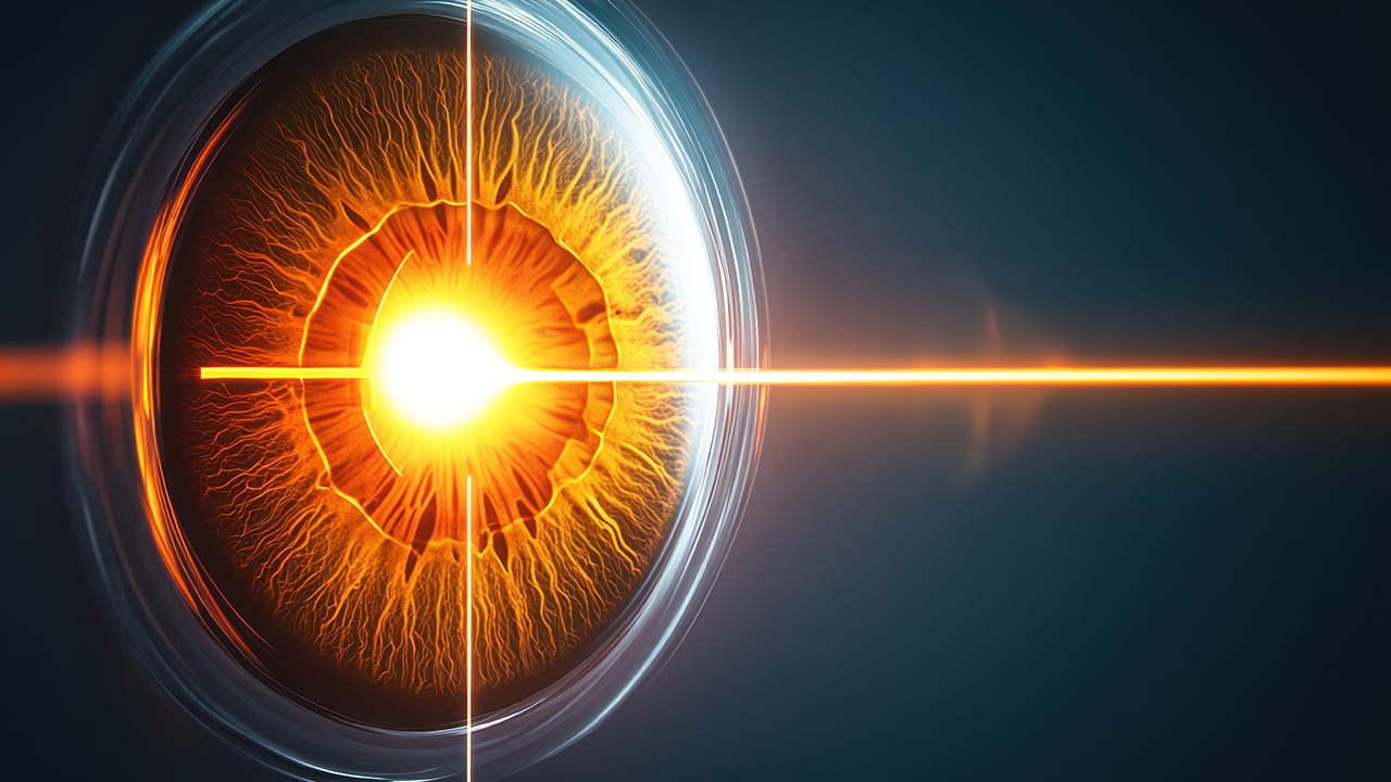
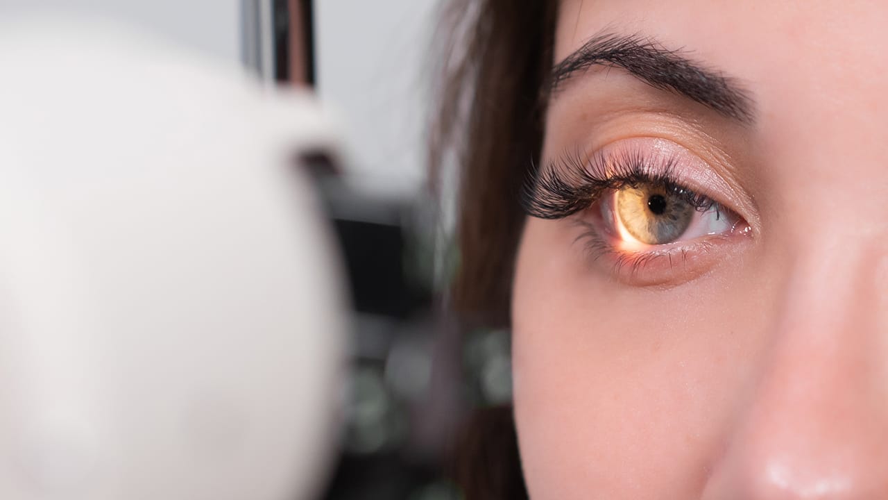
Hyperopia, also known as nearsightedness among the public, is one of the refractive errors that make it difficult to focus on near objects. If the eyeball is shorter than normal or the cornea-lens system does not refract light sufficiently to the retina, the image is formed behind the retina, not on the retina; as a result, text and details cannot be distinguished up close, while farsightedness is relatively preserved.
Hyperopia is physiologically common in infants; although it disappears in most children as they grow up, its permanent form becomes a vision problem in adulthood. Typical complaints are blurred vision up close, headache, eye fatigue and difficulty focusing in the evening. Diagnosis is made with autorefractometry and cycloplegic tests that objectively measure the refractive value. Treatment options include glasses, contact lenses and modern surgical methods.
Permanent clarity can be achieved by reshaping the cornea with excimer or femtosecond laser platforms. A correctly planned hyperopia treatment improves the quality of life, increases reading comfort and work efficiency. Regular check-ups, corneal mapping and a personalized approach are the key factors determining the success of treatment.
İçindekiler
Toggle
What is Hyperopia?
Hyperopia is a vision defect known as “hyperopia” in medical literature and causes difficulty in seeing up close. If the eyeball is shorter than normal or the cornea-lens system cannot sufficiently refract the incoming light onto the retina, the image is focused a few millimeters behind the retina, not on the retina. This simple geometric shift forces the person to see near objects blurrily; however, distant vision is usually clearer.
In hyperopia, the eye constantly works the lens and ciliary muscles to sharpen nearby details. This compensation works in the beginning; however, the effort that continues throughout the day causes headaches, eye fatigue and occasional blurred vision attacks. As the lens in the hyperopia’s eyes loses its elasticity, focus support decreases and complaints become more pronounced.
The condition can be congenital; because it is one of the most obvious types of hereditary refractive errors. If there is a family history, hyperopia is frequently seen at a physiological level in babies. In most babies, the axis length increases during the growth process, but early intervention is of great importance in children who develop permanent defects.
Hyperopia is not just a need for “reading glasses”; it can pave the way for migraine-like pain due to chronic focusing effort. Therefore, anyone who has problems with near acuity should have their refractive value measured by a specialist ophthalmologist.
What Causes Hyperopia?
Hyperopia is a vision defect that causes difficulty in near tasks, and its basis lies in differences in the anatomical dimensions of the eye. If the eyeball is shorter than normal, the cornea-lens system cannot bend the incoming light sufficiently to the retina; the focus remains a few millimeters behind the retina. This causes the person to start seeing near things blurry, while the ability to see far is relatively preserved.
Hereditary predisposition is the first risk factor. If the parents have refractive errors, it can occur physiologically in the form of hyperopia in babies; if the axis length does not increase during growth, a permanent defect develops. The structural features of the crystalline lens in the eye are also decisive; when the lens in the eyes of hyperopia cannot reach the expected refractive level, the image gathers behind the retina and the complaint of blurred vision becomes permanent.
Metabolic diseases can affect the refractive index by changing the water-protein balance of the lens. Diabetes, hyperthyroidism and long-term steroid use impair lens transparency; implantation of intraocular lenses with lower power than necessary during cataract surgery or lens displacement after trauma also trigger secondary hyperopia nearsightedness problems.
Understanding this refractive error depends on detailed biometric measurements and precise calculation of intraocular lens values. Regular check-ups, especially when diagnosed in childhood, reduce the risk of amblyopia and increase the success of future treatment alternatives.
How Does Hyperopia See?
Since hyperopia is a vision problem that makes it difficult to focus at close range, the patient experiences the most difficulty in detailed tasks such as reading, using the phone or using a needle and thread. Hyperopia constantly curves the intraocular lens to be able to see letters at a close distance; however, this temporary effort soon becomes tiring. The image that comes at rest does not fall in front of the eye but a few millimeters behind, that is, behind the retina.
The eye axis length is short in hyperopia; since the cornea-lens system does not refract the rays sufficiently, the person begins to see near objects blurry. Nevertheless, since far vision remains satisfactory at low degrees, the defect may not be noticed for a long time. In the evening hours, writings become shadowed, headaches and glare become apparent.
In order to provide image clarity, the lens in the eyes of hyperopia constantly thickens with the support of the ciliary muscle; this physiological support is strong in childhood and decreases in adulthood. Therefore, in advanced diopters, far objects can also become blurry and blurred vision spreads to all distances.
Especially high-degree hyperopia may experience loss of contrast in the dark; because when the pupil dilates, the focus error of peripheral rays increases. This situation can reduce daily comfort and direct the person to a detailed examination by the eye doctor.
Hyperopia Symptoms

Hyperopia symptoms usually appear slowly and at first the patient thinks that it is a vision problem that is only felt at the end of tiring workdays. However, this is a vision defect that causes a hidden focus effort in near vision; the lens in the eyes of hyperopia constantly thickens and tries to pull the image in front of the eye. This uninterrupted work of the muscles reveals itself with blurred vision attacks, especially in front of the screen or in long reading sessions.
-Letters appear shadowy, double or flickering at close range; the person feels the need to move the text at arm’s length.
-Dazzle in bright light, loss of contrast in dim environments are evident; tables become blurry in the evening hours.
-Throbbing headaches in the forehead and temples in adults, and rapid fatigue during lessons in children are noticeable.
-Excessive use of the eye muscles causes redness, a stinging sensation and frequent blinking reflexes.
Symptoms are also described as starting to see blurry up close, increasing eye fatigue as you read and being disturbed by strong light sources in the dark. In high-degree defects, even distant objects may lose their clarity; because the focus is still behind the retina, not on it. In case of recurrence of complaints, having an individual refractive measurement at the eye doctor without delay is critical for the success of the treatment.
How is Hyperopia Prevented?
If hyperopia is congenital, it cannot be completely prevented; however, it is possible to reduce risk factors.
– A diet rich in lutein, zeaxanthin and omega-three fatty acids
– Relaxing accommodation by looking out the window every twenty minutes in front of the screen
– Using sunglasses that protect from UV light
– Preferring homogeneous ambient light instead of dazzling lighting
Diagnosis of Hyperopia
The first step in reaching a correct diagnosis for someone who has started to see blurry up close is a detailed refraction examination. This examination begins with an automatic refractometer measurement at the eye doctor; the device analyzes the refractive environments and determines at which diopter the additional lens in front of the eye is required.
When values are high, confirmation is made with retinoscopy and subjective reading tests. The aim is to determine the most accurate plus lens power that will focus light on the retina. This process is indispensable for “hyperopic” patients who have difficulty in sharpening nearby details.
Cycloplegic drops—especially in examinations of children and adolescents—temporarily disable accommodation and reveal hidden diopters. In infants, red reflex testing and flashlight retinoscopy are critical for early detection of amblyopia risk in hyperopia screening, because the eye axis length is short in hyperopia and the image is usually focused behind the retina.
Keratometry and ultrasound biometry support the diagnosis by precisely measuring the axis length and corneal curvature. In adult patients, the cornea-lens relationship is examined in detail using optical coherence tomography or a Scheimpflug camera; these data clarify the degree of hyperopia and possible combinations (astigmatism, presbyopia) in the classification of “refractive errors”. The results serve as a road map for choosing glasses, contact lenses or surgical methods to be included in the treatment plan.
Hyperopia Test
Hyperopia test consists of visual acuity tables, retinoscopy and keratometry measurements. Cycloplegia is considered a critical step to determine the correct diopter, especially in children. The tests are non-invasive and can be completed in a few minutes; the results determine the treatment algorithm.
How is Hyperopia Treated?
Hyperopia treatment, the approach to correcting the problem of nearsightedness, is evaluated in three main steps: glasses, contact lenses and surgical methods. The aim is to focus the image on the retina by carrying the refracted light to the retina and thus provide permanent clarity.

The first step in hyperopia treatment is convex (plus) glasses with low diopters. These simple optical tools reduce eye focus effort and eliminate muscle fatigue in hyperopia. Soft silicone hydrogel contact lenses offer aesthetic and wide field of vision for patients with active lifestyles; however, they may pose a risk of infection if hygiene rules are not strictly followed.
Refractive surgery methods come to the fore in those seeking a permanent solution for hyperopia. LASIK, PRK and SMILE techniques performed with excimer or femtosecond laser platforms increase refractive power by removing tissue at micron level from the corneal surface. In individuals with limited corneal thickness, phakic IOL implantation without changing the intraocular lens gives effective results in high diopters. In patients aged 50 and over, transparent lens exchange (refractive lens exchange) may be preferred; the natural lens is removed and an artificial lens with appropriate power is placed, eliminating the risk of future cataracts.
Since refractive errors progress differently in each individual, a personalized treatment plan is essential. Therefore, detailed topography, pachymetry and biometric measurements are made before the decision for surgery. Regular follow-up examinations during the healing process play a decisive role in terms of permanent clarity and eye health.
Eye Laser Surgery for Hyperopia Treatment
LASIK (Laser-Assisted in Situ Keratomileusis)
The most frequently chosen of these surgical methods is the creation of a thin flap in the cornea with a femtosecond laser. After the flap is removed, the Excimer energy reshapes the stroma; thus, the refractive power of the cornea is increased to bend the incoming light towards the retina. The image now converges directly on the retina, eliminating the need to add convex glass to the front of the eye. The recovery rate is fast; most people no longer have blurred vision when reading a book the next day.
PRK (Photorefractive Keratectomy)
The epithelial layer of the cornea is gently lifted, and the Excimer laser is applied directly to the stroma. It is safe for patients with thin corneas or those at risk of trauma. The epithelium renews itself in three to four days, and a slight blurry vision may be experienced; however, as the defect improves, the need to use the eye muscles more in hyperopia decreases.
LASEK (Laser Epithelial Keratomileusis)
Similar to PRK; the difference is that the epithelium is softened with an alcohol solution, laid aside and covered after the procedure. This technique minimizes nerve incision and shortens the duration of pain. Not cutting the cornea preserves surface stability in the long term and helps the image to remain fixed on the retina.
SMILE (Small-Incision Lenticule Extraction)
A thin lenticule is created in the cornea with a femtosecond laser; the lenticule is extracted from only a three-millimeter incision. Since it does not require a flap, the nerve network is largely preserved and the risk of dry eyes is lower. The small incision maintains the integrity of the natural tissue at a high level after the refractive errors are corrected.
All these laser procedures reshape the cornea at the micron level to provide permanent clarity; so the lens in the eyes of hyperopia no longer has to make a constant focus effort. The specialist decides on the appropriate method after detailed topography and pachymetry; correct patient selection is the key to long-lasting results and comfort.
Frequently Asked Questions
Will my hyperopia degree increase over time?
The eye axis stabilizes in adulthood; however, presbyopic changes may reduce reading comfort.
Will the number return after hyperopia treatment?
The surgical success rate is high; rarely, low diopters may remain, this can be corrected with additional procedures.
Should I prefer contact lenses or laser?
Patients with appropriate corneal thickness who do not want to undertake the burden of daily care provide long-term comfort with laser.
Is hyperopia important in babies?
High degrees increase the risk of amblyopia; early use of glasses protects visual development.
At what age can laser be performed?
Eighteen years of age is the lower limit for refractive stability; The upper limit is related to corneal health.
Does it cause complaints other than near vision?
If the focus effort lasts long, headache and fatigue are added to the picture; school-age children may lose motivation for lessons.